
Shortwave diathermy: History and How It Works
History of heat treatment of cancer
Heat therapy, known formally as hyperthermia treatment, was used in ancient Chinese medicine, Ayurvedic medicine and in Egypt, described in papyrus on surgical technique, 5000 years ago.[1] In the time of the ancient Greeks, heat was applied to malignant tumors. Hippocrates mentions use of it in treatment of a breast tumor.
American physician William Coley reported in a landmark paper in 1893 the spontaneous regression of tumors in ten patients with bacterial induced fevers. It has been debated whether the bacterial infection or the fever induced the regression of the tumors.[2][3] Dr. Coley had observed that the patients’ tumors spontaneously reduced in size after having contracted a Streptococcus infection known at the time as erysipelas. This particular treatment method is of course considered imprudent in our time.
In 1971, American scientists Westra and Dewey wrote of their experiments in hyperthermia in mammals, and their work was the first to earn the attention of the radiation biology community of the time. In 1975, 100 participants gathered in the International Symposium on Cancer Therapy by Hyperthermia and Radiation. More international symposia followed, and the North American Hyperthermia Society was founded in 1981, later becoming the Society for Thermal Medicine.
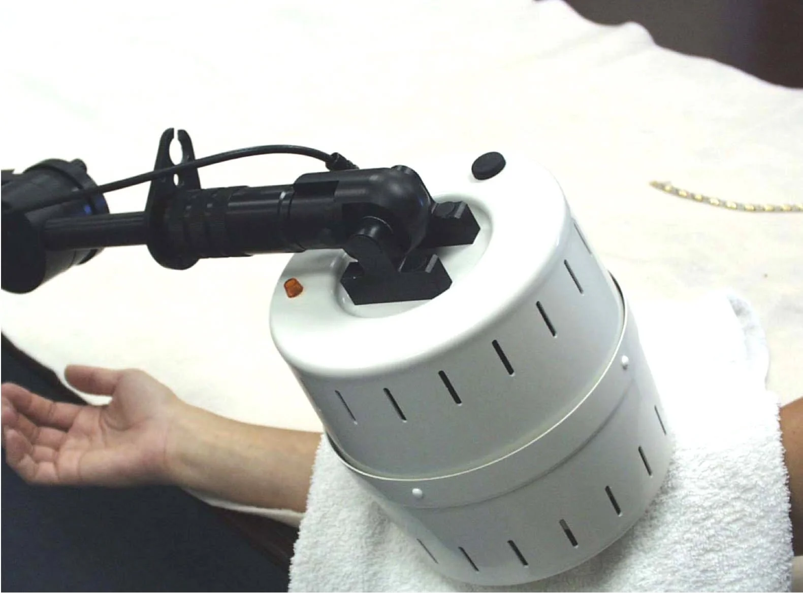
In U.S. clinics, heat therapy is used for arthritis patients, cancer patients, and painful conditions as an alternative to pain medication.
Exploiting the differences between cancer cells and normal cells
Temperatures above 122 degrees Fahrenheit (50 degrees Celsius) have been used to ablate tumor cells.[4] However, clinically, it has been observed that temperatures between 108.5 and 113 degrees Fahrenheit have killed cancer cells while leaving normal cells unharmed. Other sources find that 104 to 113 Fahrenheit (which is 40 to 44 degrees Celsius) is adequate to kill cancer cells.[5] Lethal effects on cancer cells have appeared within ten to sixty minutes.[6]
However, most normal (non-cancerous) cells were shown to be unharmed during over an hour exposure to 44 degrees Celsius (111.2 degrees Fahrenheit). Unfortunately, this study was removed from the internet, and only the abstract remains.[7] Only neural tissue was found to be more sensitive, and should not be subject to such long exposure at that temperature.
Mechanism of action
Now we should ask, given its long use throughout medical history: How does heat therapy work?
It is thought that several peculiar vulnerabilities of cancer result in the attack by way of heat that works against cancer. One such vulnerability is that cancer cells grow so quickly that they cannot acquire enough fuel for metabolism in normal temperature, let alone the increased metabolism that is stimulated by heat. The chaotic and frenzied growth of cancer cells is what renders them vulnerable to the even further extremes of hypermetabolism that targeted heat causes. As a result of heat application, we see changes in the cell membrane and nucleus, denaturation of proteins and changes in the calcium permeability in the calcium channels.[8][9] Resulting changes in the cytoskeleton and membranes overwhelm the heated cancer cells.
At first glance, this process sounds destructive. How can it be helpful to cancer patients while fighting against the cancer process?
The reason that heat is so helpful to the cancer patient, and so destructive against cancer is that there are more vulnerabilities of cancer in the presence of heat than vulnerabilities of normal tissue. Even better, there is selectivity of malignant tissue over normal in heat therapy. Heated tissue to the above temperatures results in 30% increase in intracellular temperature above normal. It so happens that malignant cells generally have rigid membranes due to increased phospholipid concentrations. So an increase in applied heat, resulting in an increase in pressure, has been found to selectively destroy malignant cells rather than normal ones,[10] particularly in the liver, brain and pancreas,[11] as well as the bladder, cervix, rectum, lung, esophagus, vulva and vagina as well as for melanoma. Those improvements were not only noted in reduction and destruction of tumor tissue, but also in overall survival from cancer.[12] Our clinic has also found heat therapy of great benefit against colon, ovarian, uterine, endometrial and prostate cancers.
Heat at the cell membrane depolarizes and destabilizes it. This uses up ATP, which is in short supply in a cancer cell, because of few and dysfunctional mitochondria to begin with, and this is why the stressed cancer cell dies. Without ATP, it cannot survive. In this way, heat exploits the vulnerabilities of cancer cells to destroy them. Normal cells, on the other hand, are resilient to the effects of moderate heat.
Cancer cells are hypoxic and are thus acidotic. This enables a further one-two punch from heat against cancer.
The hypoxia causes the cancerous tissue to rise to a higher temperature than normal tissue. Acidity of cancerous cells also was a factor in cancer’s vulnerability to heat. With pH below 7.4, exposure of tumor cells to heat increased apoptosis, especially cells in S phase, by more than a factor of 100. Hypoxia is a factor in cancer’s resistance to chemotherapy and radiotherapy, and the more hypoxic, the more death of cancer cells was found. These features, acidity and hypoxia, are both characteristics of cancer cells, and both are vulnerabilities of cancer cells to the effects of heat.
Although heat increases blood flow, it inhibits angiogenesis, by destruction of, and inability to form, new blood vessels.[13][14] This is yet another means of attack on cancer by use of heat.
Heat shock proteins are how cancer waves flags of surrender
Yet another very helpful effect of heat against cancer are heat shock proteins. These are distress signal proteins that cancer cells display on their surface after heat is applied to them. But normal cells do not express this.
The “holy grail” of cancer treatment is to find and use those treatments
that are toxic to cancer cells
while being harmless or even nourishing to normal cells.
Heat shock proteins are one such tool. The cells that expressed heat shock proteins on their surfaces were found to be more susceptible to destruction by lysis from natural killer effector cells.[15][16] And then, as a further result of the heat shock protein display, macrophages and dendritic cells join in the coordinated destruction of cancer cells.[17]
That is, in a way, heat applied to cancer cells causes those cells to wave obvious flags of surrender to the allied forces of the immune system (NK cells, macrophages and dendritic cells).
And then the destruction of the cancer cells begins.
-
E Smith. Egyptian surgical papyrus dated around 3000 BC (cited by J van der Zee: Heating the patient: a promising approach? 2002. Ann Oncol. 13. 1173-1184 ) https://www.annalsofoncology.org/article/S0923-7534(19)63846-9/fulltext
W Coley. The treatment of mailignant tumors by repeated inoculations of erysipelas. With a report of ten original cases. 1893. https://pubmed.ncbi.nlm.nih.gov/1984929/
E McCarthy. The toxins of William B. Coley and the treatment of bone and soft-tissue sarcomas. 2006. Iowa Orthop J. 26. 154-158. https://pmc.ncbi.nlm.nih.gov/articles/PMC1888599/
H Kok, E Cressman, et al. Heating technology for malignant tumors: a review. 2020. Int J Hyperthermia. 37 (1). 711-741. https://pubmed.ncbi.nlm.nih.gov/32579419/
J van der Zee. Healing the patient: A promising approach? Auf 2002. Ann. Oncol. 13 (8). 1173-1184. https://www.annalsofoncology.org/article/S0923-7534(19)63846-9/fulltext
B Giovanella, J Stehlin, et al. Selective lethal effect of supranormal temperatures on human neoplastic cells. Nov 1976. Cancer Res. 36 (11), Pt 1. 3944-3950. https://pubmed.ncbi.nlm.nih.gov/975042/
L Fajardo. Pathological effects of hyperthermia in normal tissues. Oct 1984. Europe PMC. https://europepmc.org/article/med/6467235
R Cowan, R O’Cearbhaill, et al. Current status and future prospects of hyperthermic intraoperative intraperitoneal chemotherapy (HIPEC) clinical trials in ovarian cancer. Aug 2018. Int J Hyperthermia.33 (5). 548-553. https://pmc.ncbi.nlm.nih.gov/articles/PMC5776684/#R20
J Hettinga, A Konings, et al. Reduction of cellular cisplatin resistance by hyperthermia – a review. Nov 1996. Int J Hyperthermia. 13 (5). 439-457. https://www.tandfonline.com/doi/abs/10.3109/02656739709023545
Galeotti T, et al. Membrane alterations in cancer cells: the role of oxy radicals. Membrane Pathology. Ann NY Academy of Sciences. 1986:488:468-480.
Szasz, et al. 52.
J van der Zee. Healing the patient: A promising approach? Auf 2002. Ann. Oncol. 13 (8). 1173-1184. https://www.annalsofoncology.org/article/S0923-7534(19)63846-9/fulltext
Vaupel P, et al. Physiological effects of hyperthermia. Recent Results in Cancer. 1987; 104:71-109.
Fajardo et al. Endothelial cells and hyperthermia. Int J Hyperthermia. 1994; 3:347-353.
G Multhoff, C Botzler, et al. A stress-inducible 72-kDa heat-shock protein (HSP72) is expressed ont eh surface of human tumor cells, but not on normal cells. Apr 1995. Int J Cancer. https://onlinelibrary.wiley.com/doi/abs/10.1002/ijc.2910610222
G Multhoff, C Botzler, et al. CD3- Large granular lymphocytes recognize a heat-inducible immunogenic determinant associated with the 72-kD heat shock protein on human sarcoma cells. Aug 1995. Blood. 86 (4). 1374-1382. https://www.sciencedirect.com/science/article/pii/S0006497120763949
S Basu, R Binder, et al. Necrotic but not apoptotic cell death releases heat shock proteins, which delivery a partial maturation signal to dendritic cells and activate the NF-kappa B pathway. Nov 2000. Int Immunol. 12 (11). 1539-1546. https://pubmed.ncbi.nlm.nih.gov/11058573/